| Microscope |
Type |
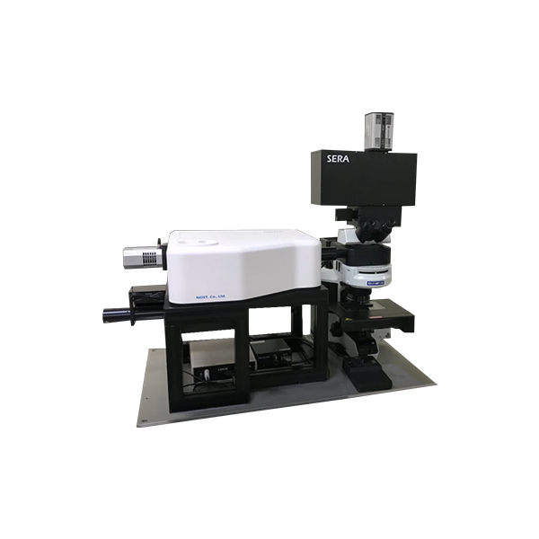
Open space research grade microscope
Upright with olympus BX5- series for opaque and transparent samples |
| Light |
white light illuminator for reflective Koehler illuminator |
| Revolver |
5 positions objective revolver with 2 objectives |
| Objective lens |
magnification : 20x ( NA = 0.45, WD = 3.1 mm)
maginification : 100x ( NA = 0.9, WD = 1 mm) |
| Video camera |
5M pixels USB color camera to visualize the image of sample |
| Binocular head |
Not included but available on request |
| Confocal |
Ture confocal and fully adjustable confocal pinhole at the spectrograph ent.
continuously adjustable from 10 to 3000 um , 10 um step |
| Microscope Enclosure |
for open lab operation (enclosure with interlock available on request) |
| Manual stage |
Travel : 70 mm x 50 mm in XY / 60 mm in Z axis
( optional large one or motorized available on request) |
| Automation |
Motorized neutral densit
y filters |
Motorized selectable 11 steps ND filter for input power adjustment |
| Shutter |
Automated mechanical shutter for preventing sample damage |
| Filter set |
Raman laser filters and PL filters controlled by SW |
| Grating turret |
Aberration corrected spectrograph with an interchangeable grating turret
(included automated calibration function) |
| laser and image |
Motorized switching between white light visualisation and
Spectrum measurement modes |
| Laser alignment |
Auto alignment and optimization of input laser
by internal reference source |
| Slit |
Motorized confocal pinhole 10 um ~ 3,000 um |
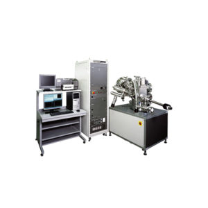
| Computer |
Suitable latest computer |
Intel core i5-7th generation, DDR4 16G RAM, VGA : 3GB |
| 1TB (SATA3 / 7200 / 64M) HDD, 250GB SSD |
| Window 7 or 10(16 BIT) |
| Monitor: FHD (1920 X 1080) |
| Software |
RAON-Spec |
Data acquisition and processing:
required accessories for instrument control, data and image acquisition, data analysis, curve plotting and Fast Mapping function |
| operating software for Window 10, including the control of
the HEDA system |
| Capabilities for both single point spectra and Raman images |
| 2D and 3D Mapping function |
| Complete automation with fully integrated spectrometer, microscope,
Detector |
| Automated spectral calibration with standard silicon |
| imaging, image analysis, storage and transfer of data in various format. |
| RAON-Vu |
Off line Viewer and analysis program |
| Open spectrum & mapping data |
| ROI analysis / Fitting ( Polynomial , Mean difference & manual)
and smoothing |
| PCA ( Principal components analysis) |
| Auto baseline correction / stray light elimination |
| Peak find / image analysis |
| Arithmetic operation |
| Create report |
| Expandability |
Hyperspectral imaging |
Hyperspectral imaging microscope to integrate with the Raman spectrome ter to provide spectral analysis of nanomaterial and image
by dark field microscope |