PHI-XPS Quantera II

X-ray Photoelectron Spectroscopy (XPS/ESCA) is the most widely used surface analysis technique and has many well
established industrial and research applications. XPS provides quantitative elemental and chemical state information from
surfaces and thin film structures. XPS is applied to a diverse range of materials applications including: polymers, metals,
catalysts, thin films, photovoltaics, batteries, wear coatings, nanomaterials, semiconductor devices, magnetic storage media,
display technology, and biomedical devices.
The Quantera II is built upon Physical Electronics’ (PHI) revolutionary scanning XPS microprobe instrument platform that
includes: a patented micro-focused scanning x-ray source, patented dual beam charge neutralization technology, a floating
column ion gun for optimized XPS sputter depth profiling, flexible robotic sample handling, and a fully automated internet
ready instrument platform. The Quantera II increases the performance and productivity of these revolutionary technologies,
providing the highest performance XPS system available to meet your current and future XPS needs.

Request Inquiry

Description

Unique Capabilities
PHI’s scanning XPS microprobe instrument platform provides secondary electron images (SXI) generated by scanning a focused 10 μm x-ray beam across the sample.
These SXI images have a contrast mechanism that is dominated by photoelectron yield (composition), and therefore often reveal features that are not visible
optically or related to topography. SXI images provide a high degree of confidence in locating small features for analysis. The micro-focused x-ray beam defines the analysis area
pattern for large area spectroscopy, micro area spectroscopy, chemical state imaging, and depth profiling.

Microprobe Workflow
A x-ray beam induced secondary electron image immediately revealed the presence of unexpected localized contaminants on a polymers surface. Micro
area spectra obtained with a 20 μm diameter x-ray beam identified a fluorocarbon contaminant in a few minutes. Elemental maps show the smaller features in
the secondary electron image do not contain fluorine. A micro area spectrum from the smaller features, obtained using a 10 μm diameter x-ray beam, identifies
the presence of a second contaminant that contains Zn.

High Productivity
Whether you are analyzing a thin polymer sheet, a large plastic lens, a steel razor blade, or electrically isolated solder bumps; the instrument set-up is the same. Point
and click at an optical image to select the analysis areas. Then start the analysis with the dual beam neutralizer and the auto-Z functions activated to provide automatic
sample alignment and charge neutralization. There is no individual sample tuning, no concerns over sample composition and size, and no worries about walking
away from the instrument and letting it automatically collect data from all your samples.

Optimized Configuration
A focused x-ray beam, high sensitivity spectrometer, high performance floating column ion gun, turnkey dual beam charge neutralization, compucentric Zalar rotation, and
advanced data reduction algorithms provide the highest performance XPS depth profiling capability available. Applications include: semiconductor thin film structures,
magnetic media thin films, optical coatings, decorative coatings, wear coatings and, with the optional C60 or GCIB cluster ion beams, polymer and organic thin films
such as time release drug coatings and organic LED films.

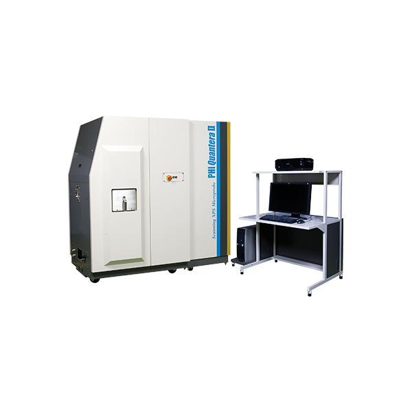
PHI Quantera II Scanning XPS Microprobe

The core technology of the PHI Quantera II is PHI’s patented, monochromatic, micro-focused, scanning x-ray source which provides excellent large area and superior micro-area spectroscopy performance. Spectroscopy, depth profiling, and imaging can all be performed over the full range of x-ray beam sizes including the minimum x-ray beam size of less than 7.5 µm. In addition to superior XPS performance characteristics the PHI Quantera II provides two in situ sample parking stations which enables the automated analysis of all three sample platens in a single user defined analysis queue.

Intuitive Sample Navigation And Confident Analysis Area Identification

  • The unique scanning X-ray microprobe allows SEM like navigation with point-and-click control
  • X-ray induced secondary electron imaging (SXI) provides perfect correlation between imaged areas and spectroscopy

Optimized Depth Profiling

  • Multiple ion gun options (monatomic Ar, C60, argon cluster GCIB) for a variety of organic, inorganic, and mixed materials
  • Full 5-axis stage functionality including rotation/tilt and heating/cooling during sputtering
  • Multipoint profiling within a single sputter crater for on/off defect analysis and precious samples
  • Adjustable solid collection angle for improved angular resolution for Angle Resolved analysis with advanced software for high-throughput film structure analysis

Superior Micro-Area Analysis

  • Highest small area sensitivity on the market
  • <10 microns microprobe size in x and y
  • Image registration for unattended automated micro-area analysis

Suite Of Specialized Solutions For Advanced Surface Analysis Needs

  • Heating and Cooling in situ
  • Electrochemical (biasing, polarization studies) experiments
  • Glove Box Adapter